
5:2 rotary motors in motility and anti-phage defence

The Zorya anti-phage defence system contains ZorAB, a membrane complex with the architecture of the flagellar stator unit. We study how Zorya detects phage infection and inactivates phage DNA.

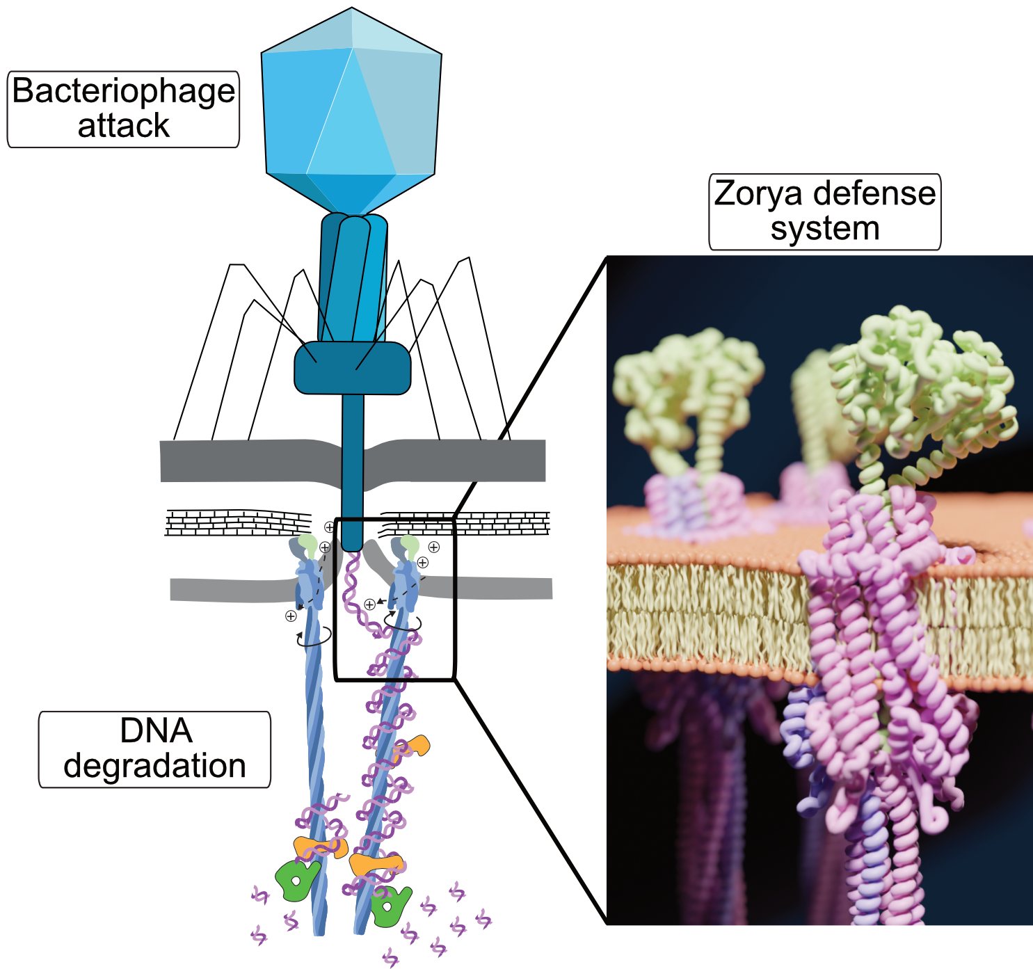
MotA5B2 and ZorA5B2 at the same scale (26 s). The function of the ZorA tail is a proposed model.
Fig. 2 of Hu, Popp et al. 2025, Nature
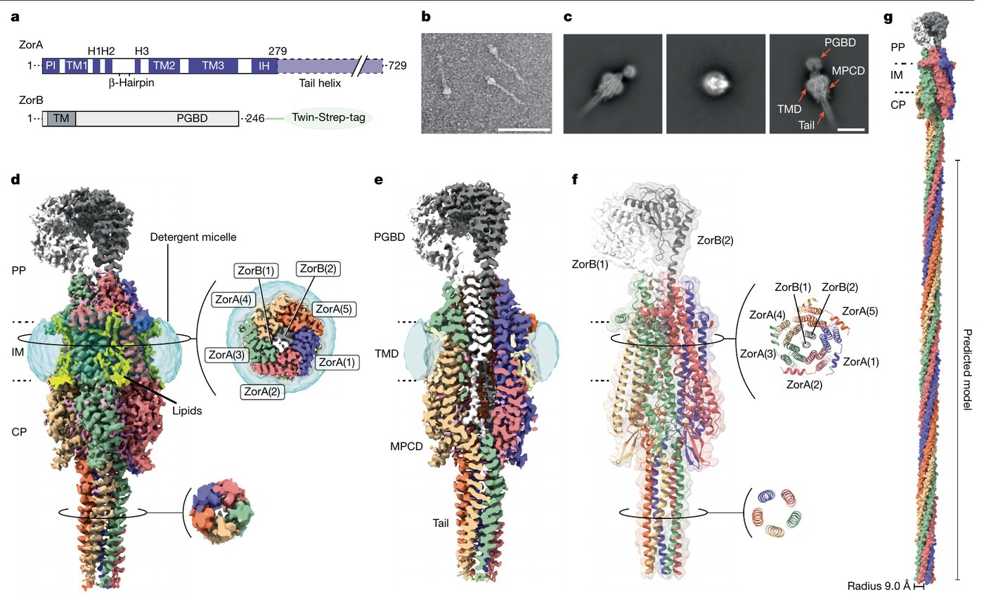
Key figure Cryo-EM structure of ZorAB. Five ZorA subunits surround two ZorB subunits; the ZorA tail extends into the cytoplasm (g, predicted model of the full-length complex). Fig. 2 of Hu, Popp et al. 2025, Nature, CC BY-NC-ND 4.0.

Bacteria encode many defence systems against bacteriophages. Zorya is widespread, and three types are known. Its membrane complex ZorAB resembles the flagellar stator unit MotAB and carries a long cytoplasmic tail.

We use our knowledge of the flagellar motor to determine what activates ZorAB and how its rotation is coupled to defence.

Findings

70 nmlength of the ZorA tail

ZorAB is a 5:2 complex with a 70 nm cytoplasmic tail

Together with the Taylor and Jackson groups, we determined cryo-EM structures of ZorAB. Five ZorA subunits surround two ZorB subunits. ZorB contains a dimeric peptidoglycan-binding domain. ZorA forms a pentameric coiled-coil tail that extends about 70 nm into the cytoplasm.

Hu et al. 2025, Nature

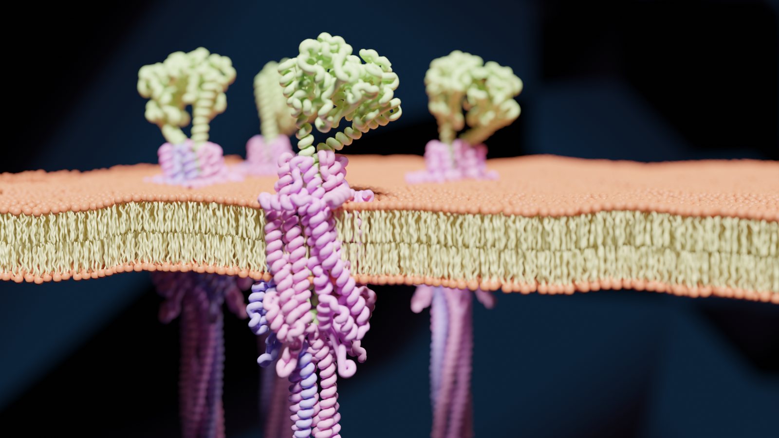
Rendering of ZorAB complexes in a membrane
ZorAB in the inner membrane, rendered from the cryo-EM structure (Hu, Popp et al. 2025).

Zorya degrades phage DNA after perturbation of the cell envelope

Mutational and functional analyses showed that all Zorya components are required for defence. ZorC binds DNA, and ZorD has nuclease activity. The data support a model in which ZorAB is a proton-driven motor that is activated when a phage perturbs the cell envelope. In this model, the ZorA tail recruits ZorC and ZorD, which degrade the phage DNA.

Hu et al. 2025, Nature

Proposed mechanism of Zorya (28 s). Winding of DNA by the ZorA tail is a hypothesis.

Druantia recognises exposed single-stranded DNA

Together with the Wahl and Beisel groups, we characterised the type III-A Druantia system of Escherichia coli. DruE forms a dimer, binds exposed single-stranded DNA and unwinds DNA in the 3′-to-5′ direction. DruE and DruH together cleared phage infections without an effect on cell growth or viability.

Himpich et al. 2026, Cell Reports

Salmonella enterica subsp. salamae encodes a contractile injection system

Together with the Taylor group, we determined the cryo-EM structure of this extracellular contractile injection system. Its sheath architecture differs from that of other contractile injection systems, and a cage-like shell surrounds the central spike. Our data indicate that it interacts with the inner membrane.

Ejaz et al. 2026, Nat Commun

Current projects

Sensing of phage infection by Zorya

DFG priority programme SPP 2330, joint project with Philipp Popp

We determine which signal activates Zorya, how its components are recruited to the infection site and how they interfere with phage DNA injection and replication. We compare Zorya types I to III in Gram-negative and Gram-positive bacteria. We use TIRF and STED microscopy, HaloTag and SNAP-tag labelling, single-molecule tracking, proximity labelling, RNA sequencing and phage screens.

Contact: Marc Erhardt, E-mail · Project page at SPP 2330

Selected publications

Next topic: Expression and substrate targeting of the SPI-1 injectisome