

Image analysis tools for single-cell microscopy

Single-cell experiments generate large time-lapse data sets. We develop software for automated segmentation, tracking and quantification of bacteria.

Time-lapse of cells in a mother machine. White: phase contrast. Red: cytoplasm. Yellow: foci. Yellow outlines: automated segmentation.
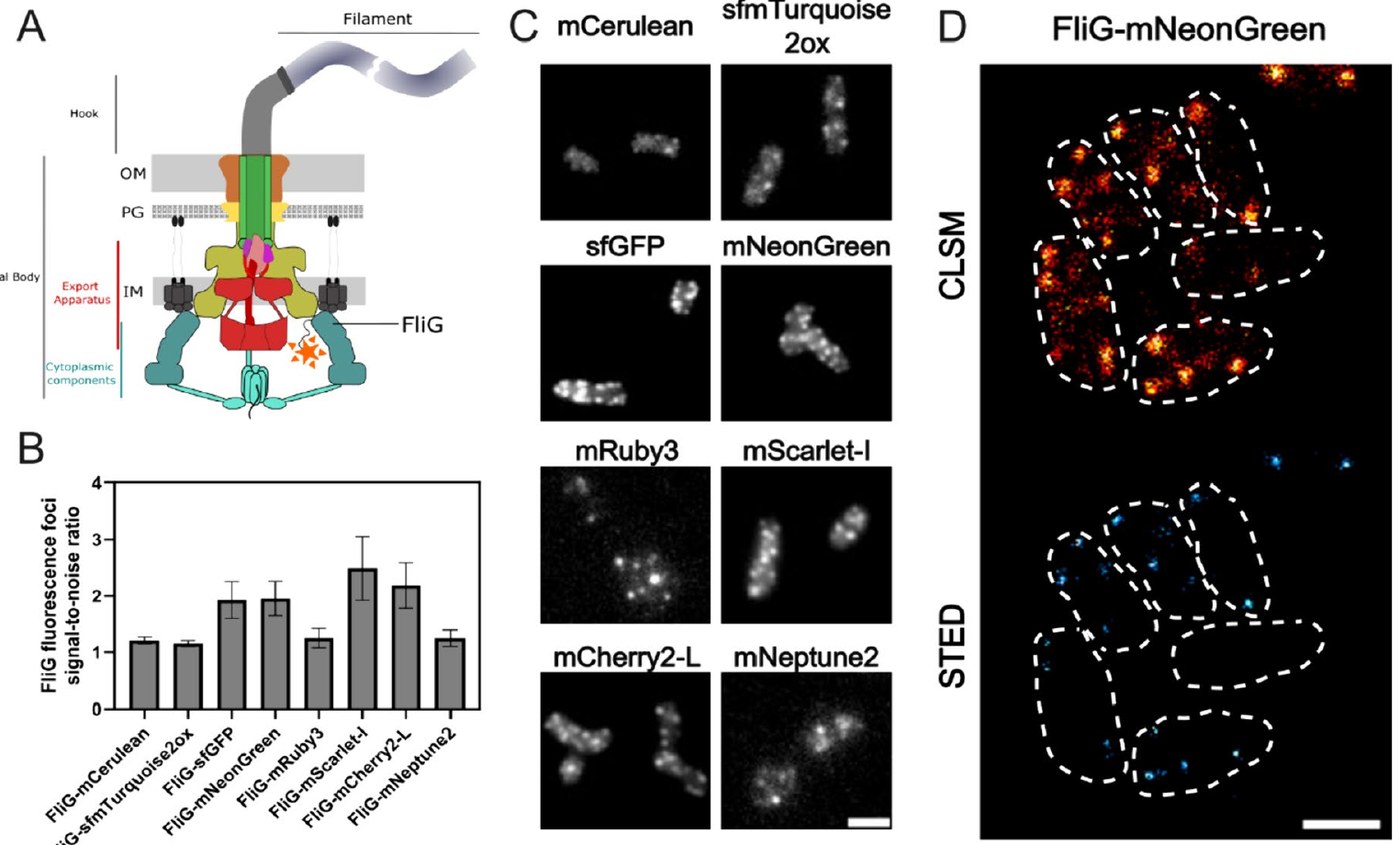
Fig. 6 of Delgadillo-Guevara et al. 2024, Journal of Biological Engineering
Key figure FliG fused to different fluorescent proteins in Salmonella: signal-to-noise ratio, epifluorescence images and confocal and STED images of FliG-mNeonGreen. Fig. 6 of Delgadillo-Guevara et al. 2024, Journal of Biological Engineering, CC BY 4.0, cropped.

Several of our projects require measurements in single cells over many generations, for example the number of flagella per cell or the localisation of a defence complex. These measurements depend on automated and reproducible image analysis.

Workflow

  1. 1Importnd2, tif and other formats, with their metadata
  2. 2Pre-processingPreparation of the images for segmentation
  3. 3Segmentationilastik, Omnipose or DeLTA
  4. 4Quality controlFilters by cell morphology
  5. 5QuantificationFluorescence, foci and growth
  6. 6Tracking and exportLineages, kymographs, structured tables

Tools

Mother machine

MMAPMother Machine Analysis Platform

MMAP analyses mother-machine experiments from the raw microscopy files to single-cell tables.

Segmentation of single cells with Omnipose.
  • Reads nd2, tif and other microscopy formats and extracts the metadata
  • Segmentation with ilastik, Omnipose or DeLTA
  • Kymographs of single channels
  • Batch processing of complete experiments

Preprint 2026

BactoMateIntegrated platform for bacterial microscopy analysis

BactoMate is an open-source, cross-platform graphical user interface that joins the steps of microbial image analysis in one workflow. We tested it on multichannel fluorescence images, swimming assays, microcolony lineages, phage infection assays and microfluidic time series.

  • Pre-processing and cell segmentation with established methods
  • Quality control by cell morphology
  • Fluorescence and foci quantification
  • Single-cell tracking and lineage reconstruction
  • Single files or batch processing
  • All parameters are saved with the results and can be loaded again

Published

Fluorescent toolsStandardised work in Gram-negative bacteria

A set of characterised fluorescent tools for Gram-negative bacteria, and a guide for membrane potential measurements with voltage-sensitive dyes (with the Strahl group).

  • FliG fused to different fluorescent proteins in Salmonella
  • Signal-to-noise ratios in epifluorescence, confocal and STED microscopy
  • Protocols for membrane potential measurements

Open-source code

All repositories of the lab on GitHub

Current projects

Development of image analysis tools

We adapt the analysis tools to new experimental designs and imaging modalities.

Contact: Lasse Hallenga, E-mail

Selected publications

Next topic: Assembly, protein export and rotation of the bacterial flagellum